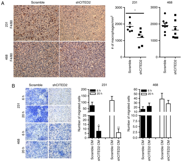
Figure 1.
Effect of silencing CITED2 on macrophage infiltration in vivo and recruitment in vitro. (A) Representative images of immunohistochemical analysis revealed staining for the macrophage marker F4/80, as indicated by 3,3-diaminobenzamindine staining (brown) and visualized using a light microscope (magnification, ×200). The adjacent histograms represent the average number of macrophages/mm2. *P<0.05 compared with the scramble group. (B) Representative images revealed macrophage recruitment in response to conditioned media obtained from scramble and shCITED2-expressing breast cancer cells at 6 and 20 h, respectively, using an in vitro Transwell migration assay (magnification, ×400). The adjacent histograms represent quantification of the average number of macrophages recruited at each time point. *P<0.05; **P<0.01 vs. scramble CM. CITED2, Cbp/p300-interacting transactivator with Glu/Asp-rich carboxy-terminal domain-2; sh, short hairpin; CM, conditioned media.

