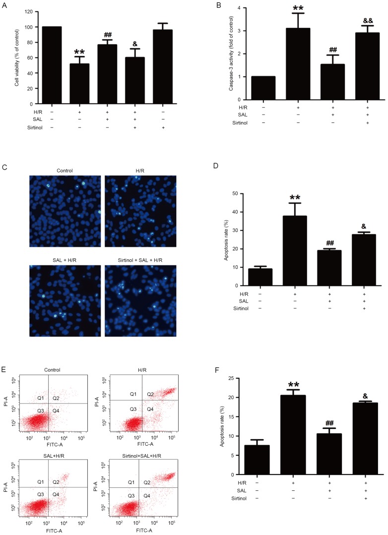
Figure 4.
Effects of sirtinol on the SAL-induced protection against H/R-induced cytotoxicity and apoptosis in HBVSMCs. Following pretreatment with sirtinol (10 µM) for 30 min, HBVSMCs were incubated with SAL (400 µM) for 30 min and then exposed to hypoxia for 8 h followed by reoxygenation for 16 h. (A) The viability of HBVSMCs was detected by MTT assay and expressed as a percentage of the control. (B) Caspase-3 activity was measured using a commercialized caspase-3 assay kit and expressed as a fold change compared with the control. The morphological changes in the apoptotic cells were observed by (C) Hoechst 33324 staining (magnification, ×200) and assessed by (D) statistical analysis of the ratio of apoptotic cells to total cells. (E) The apoptosis ratio of HBVSMC was detected by Annexin V-FITC/PI staining and assessed by (F) statistical analysis. Data are presented as the mean ± standard error of the mean from three independent experiments performed in triplicate. **P<0.01 vs. control group; ##P<0.01 vs. H/R group, &P<0.05, &&P<0.01 vs. SAL and H/R co-treatment group. SAL, salidroside; H/R, hypoxia/reoxygenation; HBVSMCs, human brain vascular smooth muscle cells; FITC, fluorescein isothiocyanate; PI, propidium iodide.

