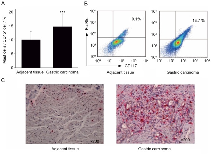
Figure 1.
Differential expression of mast cells in gastric cancer and adjacent tissues. (A) The proportion of mast cells was significantly increased in gastric cancer, compared with adjacent tissue. (B) Flow cytometry and (C) immunohistochemical analysis validated that the expression of mast cells in gastric cancer tissues was increased, compared with adjacent tissue mast cells (×200 magnification). Representative images are presented. ***P<0.001 vs. adjacent tissue. CD, cluster of differentiation; FcεRIα, high-affinity immunoglobulin E receptor.

