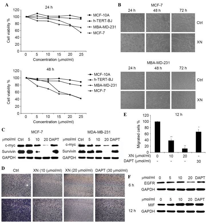
Figure 2.
XN inhibited breast cancer cell proliferation in a dose- and time-dependent manner. (A) h-TERT-BJ, MCF-10A, MCF-7 and MDA-MB-231 were plated equally (six replicates) and treated with XN. MTT assays were performed daily for two days. DMSO was a vehicle control. (B) MDA-MB-231 and MCF-7 cells were treated with 20 µM XN for 24–72 h and visualized by light microscopy. (C) The c-Myc and survivin protein expression levels were detected by western blotting. (D) The Boyden chamber transwell assay demonstrated MDA-MB-231 cell migration. MCF-7 and MDA-MB-231 cells were treated with XN (data are presented as the mean ± SEM of three independent experiments). (E) Quantification of migrated MDA-MB-231 cells. Data are presented as the mean ± SEM of three independent experiments. *P<0.05, **P<0.01 vs. control. (F) EGFR and MIF expression levels were determined by western blot analysis. SEM; standard error of the mean; DMSO, dimethyl sulfoxide; EGFR, epidermal growth factor receptor; MIF, tumor metastasis-associated protein; CDK4, cyclin-dependent kinase 4; DAPT, Duration of Dual Antiplatelet Therapy; XN, xanthohumol; Ctrl, control.

