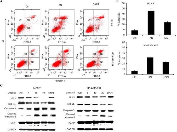
Figure 4.
XN promote apoptosis of breast cells. (A) MCF-7 and MDA-MB-231 cells were harvested for apoptotic analysis using Annexin V-FITC staining. (B) Quantification of apoptosis. Data are presented as the mean ± SD error of the mean of three independent experiments. (C) Apoptosis associated proteins Bcl-2, Bcl-xL and caspase-3 and cleaved PARP were analyzed by western blotting. GAPDH was used as the control. FITC, fluorescein isothiocyanate; FITC-A, FITC-annexin; Ctrl, control; XN, xanthohumol; DAPT, Duration of Dual Antiplatelet Therapy; PI-A, propidium iodide annexin; Bcl-2, B cell lymphoma-2; Bcl-xL, B cell lymphoma extra 1; PARP, poly (ADP-ribose) polymerase.

