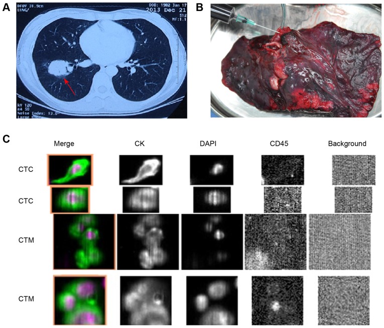
Figure 1.
Blood collection and identification of CTCs and CTM. (A) Patient with a tumor in the right lower lobe of the lung (red arrow). (B) Upon lobectomy, a syringe was inserted into the pulmonary vein, and a 7.5-ml blood sample was immediately collected. (C) Tumor cells were defined as those merged fluorescent images (from two patients) with round to oval morphology containing DAPI-stained nuclei and expressing CKs but not CD45. CTM were identified as CTC clusters containing ≥3 nuclei (magnification, ×100). CK, cytokeratin; CTC, circulating tumor cell; CTM, circulating tumor microemboli; CD45, cluster of differentiation 45.

