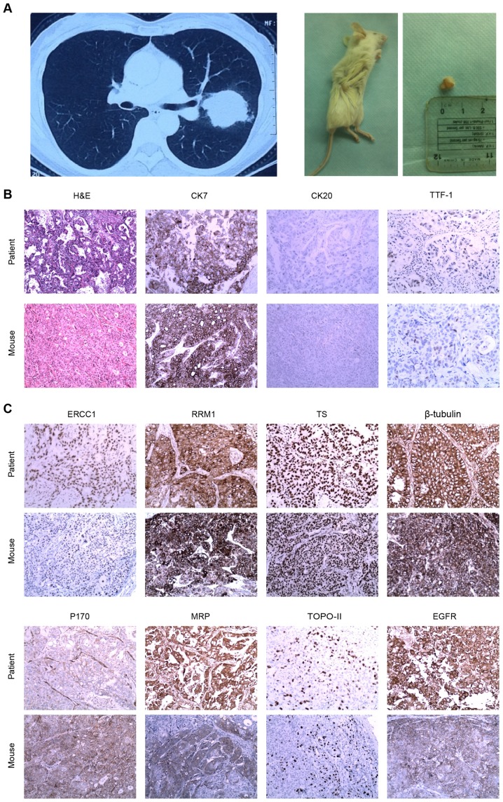
Figure 4.
Xenograft assay and IHC staining. (A) Patient with a tumor in the left upper lobe of the lung (left panel) and mouse xenograft tumor formed following injection with circulating tumor cells from the pulmonary vein of the patient (right panel). (B) H&E and CK7-positive, CK20-negative and TTF-1-positive IHC staining of the primary tumor from the patient and the mouse xenograft tumor. (C) IHC staining of a series of drug resistance-associated proteins in the primary and xenograft tumors (magnification, ×20). H&E, hematoxylin and eosin; CK, cytokeratin; TTF-1, thyroid nuclear factor-1; ERCC1, excision repair cross-complementing 1; RRM1, ribonucleotide reductase M1; TS, thymidylate synthase; MRP, multi-drug resistance-associated protein; TOPO-II, topoisomerase II; EGFR, epidermal growth factor receptor; IHC, immunohistochemistry.

