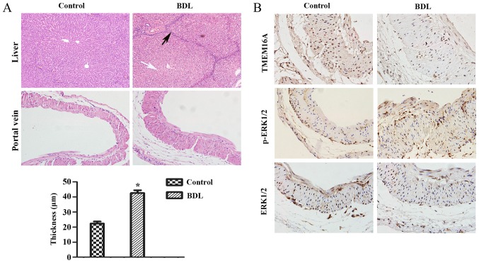
Figure 1.
Pathological alterations and protein expression levels in the rat cirrhotic liver at 8 weeks after BDL. (A) The extent of liver fibrosis (magnification, ×200) and the wall thickness of the portal vein (magnification, ×400) were assessed by hematoxylin and eosin staining. Data are presented as the mean ± standard deviation (n=10). Compared with the normal structure of liver, severe degeneration associated with necrosis were observed in model group (white arrow), accompanied by inflammatory infiltration around the portal area, a wide range of hyperplasia in connective tissues and destruction in lobular structure (black arrow). Compared with the control group, the thickness of the portal vein in the model group was increased (21.75±5.56 µm vs. 43.27±9.62 µm). *P<0.05 vs. control group. (B) Protein expression levels of TMEM16A, p-ERK1/2, ERK1/2 in the portal vein were visualized by immunohistochemistry. BDL, bile duct ligation; TMEM16A, transmembrane protein 16A; ERK1/2, extracellular signal-related kinase 1 and 2; p-ERK1/2, phosphorylated ERK1/2.

