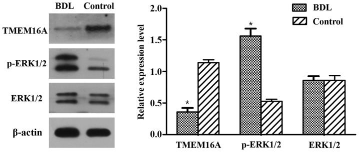
Figure 2.
Protein expression levels of TMEM16A, ERK1/2 and p-ERK1/2 in BDL rats. Protein expression was detected by western blotting of portal vein sections isolated from BDL and control rats. Protein expression levels were normalized against β-actin. Experiments were performed in triplicate. Data are presented as the mean ± standard deviation. *P<0.05 vs. control. BDL, bile duct ligation; TMEM16A, transmembrane protein 16A; ERK1/2, extracellular signal-related kinase 1 and 2; p-ERK1/2, phosphorylated ERK1/2.

