
Fig. 1.
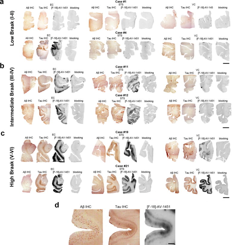
Microphotographs of postmortem brain tissue sections depicting Aβ and PHF-1 tau immunostaining, [F-18]-AV-1451 phosphor-screen autoradiography and blocking conditions in adjacent slides from representative subjects at different Braak stages [2]: (a) low Braak, (b) intermediate Braak, and (c) high Braak, and high magnification microphotographs depicting Aβ and PHF-1 tau immunostaining and [F-18]-AV-1451 phosphor-screen autoradiography of the STS from case #21 (d). [F-18]-AV-1451 binding was detected in all tangle-containing regions matching the pattern of PHF-tau immunostaining across the different Braak stages, but not the Aβ immunosignal. No [F-18]-AV-1451 binding was detected in white matter or in cortical regions lacking NFTs. Scale bar: 1 cm (a-c), 2 mm (d). Abbreviations: Aβ = β-amyloid; IHC = immunohistochemistry; NFT = neurofibrillary tangles; PHF = paired helical filament