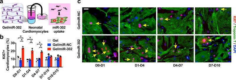
Figure 2. In vitro cardiomyocyte proliferation.
a) Schematic of miR-302 supernatant collection and cardiomyocyte uptake. Gel/miR-302 (100 µL) assemblies were formed in 1.5 mL microcentrifuge tubes with cholesterol-modified miR-302 (210 µM of miR-302b and miR-302c) or miR-NC (420 µM). OPTI-MEM (500 µL) was added above gels and supernatant was collected, frozen, and replaced at D1, D4, D7, D10 and D15. Supernatants collected from each timepoint were added to primary neonatal cardiomyocytes in culture. At 48 hours, cardiomyocytes were stained for Ki67, cardiac Troponin T, and with DAPI to detect proliferation. b) Quantification of Ki67+cTnT+ neonatal cardiomyocytes from gel supernatants from D0-D1, D1-D4, D4-D7, D7-D10 and D10-D15 in vitro cultures demonstrating proliferative effects from early gel/miR-302 release out to 7 days (mean ± SD, n=3 per condition, *p<0.05). Quantification was based on counting of Ki67+cTnT+ co-stained cells relative to total cTnT+ cells per high power field (HPF). c) Representative images of Ki67+cTnT+ neonatal cardiomyocytes (yellow arrows) to demonstrate increased Ki67 staining up to 7 days after exposure to gel/miR-302. Scale bar: 25 µm.

