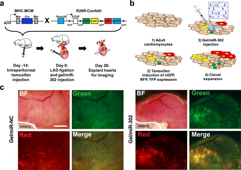
Figure 4. Gel/miR-302 induced clonal expansion in vivo.
a) Schematic representation of lineage-tracing strategy and experimental design. To trace clonal proliferation, mice were cross-bred with Myh6MerCreMer and R26RConfetti. The expression of Myh6 leads to Cre-loxP recombination with consequent random activation of one of four fluorescent reporter proteins (nGFP, YFP, RFP and mCFP) with each color representing a different clone from a Myh6 positive cardiomyocyte. In our experimental design, mice were injected with tamoxifen intraperitoneally to induce the stochastic expression of nGFP, RFP, YFP and mCFP. After 14 days, the LAD was ligated to induce ischemic injury and gels were injected in the border zone of the infarct downstream. At 28 days after ligation, hearts were collected for analysis of clonal expansion. b) Mechanism for gel/miR-302 induced clonal expansion: 1) adult cardiomyocytes are non-proliferative and incapable of dividing after ischemic injury; 2) tamoxifen is used to randomly label a small population of cardiomyocytes with one of four fluorescent reporter proteins; 3) hearts are infarcted and gel/miR-302 is injected into cardiomyocytes in the border zone; and 4) miR-302 stimulates differentiation, proliferation, and expansion of fluorescently-labeled cardiomyocytes, which pass the fluorescent protein gene onto daughter cells. c) Fluorescent scans of gross heart specimens immediately after explanting at 28 days. Fluorescence is displayed in the green and red channels to indicate labeling of cardiomyocytes in both gel/miR-NC and gel/miR-302 treated groups. Scale bar = 2 mm.

