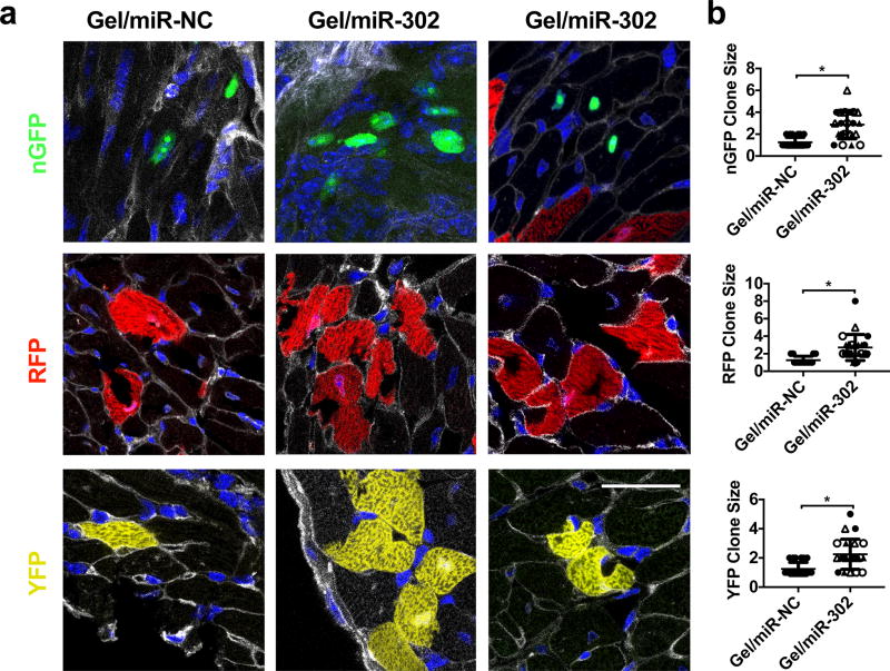
Figure 5. Clonal expansion of Confetti-labeled cardiomyocytes in mice.
a) Representative sections from confocal imaging with labeled cardiomyocytes expressing nGFP, RFP, or YFP. Gel/miR-NC sections consisted mostly of individual cardiomyocytes that were spatially separated. In gel/miR-302 treated groups, multiple clones were observed in all three fluorescent channels in close proximity, consisting of several daughter cells from a single parent cell. WGA separates individual cardiomyocytes and permits identification of clones, specifically to differentiate multiple cardiomyocytes from multi-nucleated cardiomyocytes. Scale bar = 50 µm. b) Quantification of cells to a clone in the nGFP, RFP, and YFP channels. Clones are identified as cells within 50 µm proximity to one another (gel/miR-NC, n=3; gel/miR-302, n=4, symbol shapes indicate each animal, *p<0.05). Clones consisting of one cell are not technically clones but stochastically labeled single cells, but are still counted as part of the analysis to demonstrate they are the ubiquitous in the gel/miR-NC groups.

