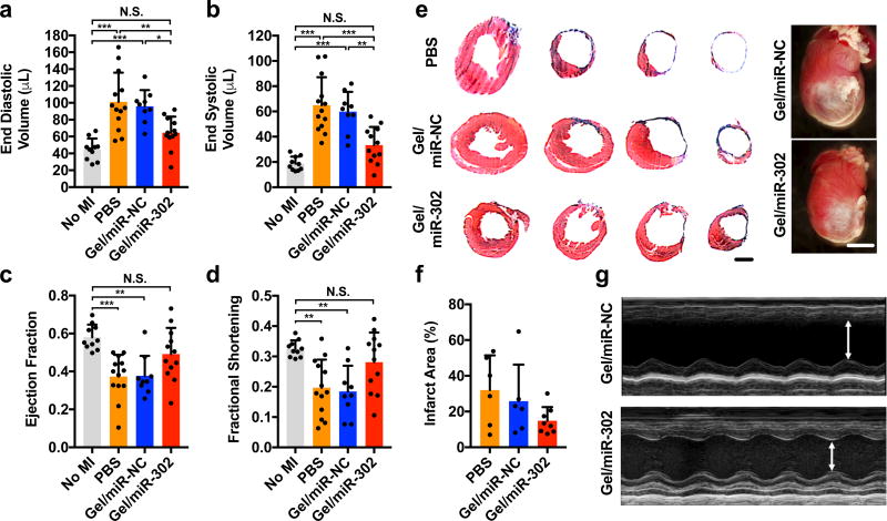
Figure 7. Functional outcomes after myocardial infarction in adult mice.
a) End diastolic and b) end systolic volumes at 4 weeks after myocardial infarction in mice treated with PBS, gel/miR-NC, or gel/miR-302 by B-mode echocardiography. Volume increases were significantly decreased in gel/miR-302 treated groups compared to controls. c) Ejection fraction and d) fractional shortening at 4 weeks after myocardial infarction by echocardiography. Neither ejection fraction nor fractional shortening of gel/miR-302 treated mice were significantly different from non-infarcted mice. All groups were compared through 1-way ANOVA (Mean ± SD, no MI, n=10; PBS, n=13; gel/miR-NC, n=9, gel/miR-302, n=12. *p<0.05 **p<0.01 ***p<0.001). Outliers were removed using the robust regression and outlier removal method (ROUT) in Prism. e) Representative Masson’s trichrome sections demonstrating cardiac volume improvement at 28 days. Sections are arranged from ligation to the apex to visualize changes in tissue remodeling. Scale bar (sections)= 2 mm. Scale bar (gross) = 5 mm. Representative gross heart pictures at D28 are also shown. f) Quantification of infarct size from gross sections. Infarct size was calculated from a minimum of three sections per animal where the scar was well-represented and expressed as Infarct Area (%) = (Infarct Area)/(Total Section Area). g) M-mode echocardiographs of left ventricular anterior and posterior walls demonstrating diminished motion in anterior wall of gel/miR-NC treated mice with improvement in gel/miR-302 treated mice.

