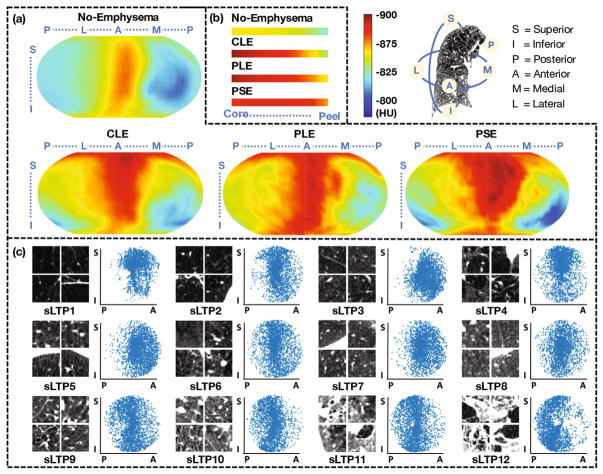
Fig. 2.
(a) Average intensity (HU) mapped onto PDCM surfaces for no-emphysema, CLE-, PLE- and PSE-predominant subjects (N = 205, 37, 12 and 10 respectively). (b) Core to peel average intensity for the same population. (c) Random ROI samples (axial cut) and sagittal spatial scatter plots of 12 sLTPs learned on the full training set.

