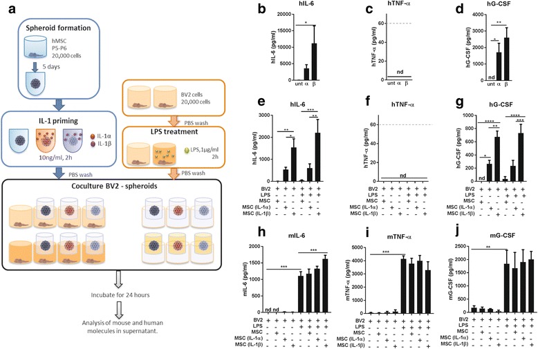
Fig. 4.

Treatment of BV2 using spheroids in inserts. a Mesenchymal stem cells (MSCs) were expanded and cultured in 3D to form spheroids, primed with interleukin (IL)-1 and co-cultured in inserts with BV2 cells previously treated with lipopolysaccharide (LPS). Measurements of human (h) cytokines on the conditioned media added to the BV2s (b–d), and in the conditioned media obtained from cells after 24 h in the inserts (e–g). Murine (m) cytokines were also analysed from the co-culture supernatants (h–j). Analysis of the supernatant of the co-culture revealed a significant increase in mIL-6 after adding conditioned media (CM) from IL-1β-primed spheroids. Dashed lines in c and f indicate the quantification limit for tumour necrosis factor (TNF)-α ELISA. *p < 0.05, **p < 0.01, ***p < 0.001, ****p < 0.0001. G-CSF granulocyte-colony stimulating factor, nd not detectable, P passage, PBS phosphate-buffered saline, unt untreated
