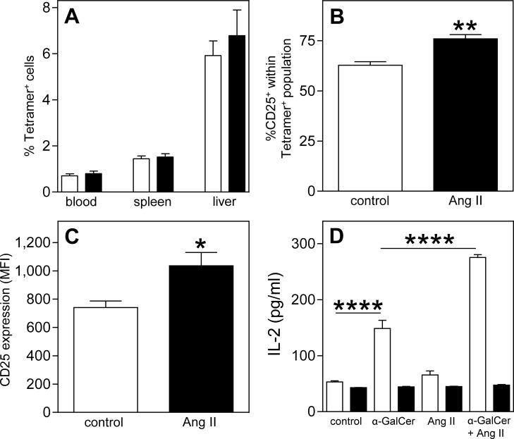
Fig 3. Increased NKT cell activity upon angiotensin II treatment.
Osmotic pumps filled with PBS (n = 5) or Ang II (n = 5) were implanted in LDLr-/- mice fed a Western type diet for 1 week. Two weeks after pump placement, the mice were sacrificed and the percentage of NKT (Tetramer+) cells in spleen and liver (A, white bars represent PBS treated mice, black bars Ang II treated mice) and the activation status of splenic NKT cells (B and C) were determined by FACS analysis. To confirm these effects, antigen-presenting cells (APCs) isolated from bone marrow of LDLr-/- (white bars) and LDLr-/-CD1d-/- mice (black bars) were incubated with α-GalCer, AngII or a combination of both. Four hours after incubation, the APCs were co-cultured with DN32.D3 hybridoma cells. After 24 hours, the IL-2 concentration in the supernatant was determined (D). All values are mean±SEM and statistical analysis was performed using the unpaired two-tailed student’s T-test (A-C) or one-way ANOVA (D) *P<0.05, **P<0.01, ****P<0.0001.

