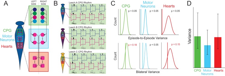
Figure 1. Schematic detailing the different components and activities within the leech heart system.
(A) The heart system of the leech consists of two symmetrical tubes that run along the left and right side of the body. Each tube has components at three levels: the central pattern generator (CPG) interneurons (green box), which connect to the motor neurons (blue box), which in turn stimulate the heart muscles (red box). (B) Wenning et al. measured various phase differences in different leeches: for example, the phase difference ΔφA1 is the first measurement of the delay between the activation of two particular neurons in the CPG in leech A, and ΔφC4 is the fourth measurement of the delay between the activation of these neurons in leech C. Measuring how particular phase differences change over time allows the episode-to-episode variance to be determined, and comparing results for the left and right sides allowed the bilateral variance to be determined. (C) Wenning et al. compared their results (vertical lines) for episode-to-episode variance (top) and bilateral variance (bottom) with theoretical predictions for a randomized population (curves). Because the measured episode-to-episode variance was significantly different from the population variance (top), there must be physiological constraints on the episode-to-episode variability at all three levels. The measured bilateral variance was also significantly different from the population variance for motor neurons, but not for the CPG and the heart muscle component, which suggests that the two sides of a single animal are no more similar to one another than two randomly chosen individuals. (D) Comparisons across all three levels revealed that the motor neurons have lower variance than the CPG or heart muscle components. Error bars reflect 95% confidence intervals.

