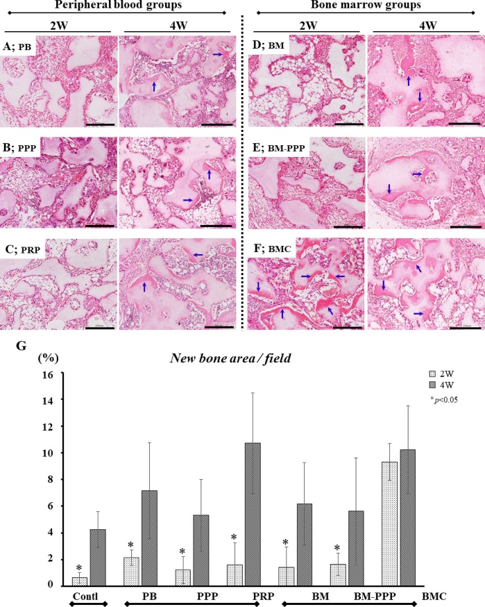
Fig 2. Typical histological appearance in specimens at 2 and 4 weeks following subcutaneous transplantation to the dorsal skin.
For each group, sections were stained with hematoxylin and eosin (H&E), and the scale bars represent 200 μm. The left and right panels show the typical appearance at 2 and 4 weeks, respectively, following transplantation. In the (A) PB, (B) PPP, (C) PRP, (D) BM, and (E) BM-PPP groups, minimal ectopic bone was detectable at 2 weeks but promoted bone formation (blue-arrows) was observed at 4 weeks. In the (F) BMC group, abundant ectopic bone (blue-arrows) was observed at 2 and 4 weeks. (G) The average area (%) of ectopic bone per whole area was measured in each group. At 2 weeks, the BMC group exhibited significantly bone formation (9.3±1.4%) compared to the other groups (approximately 1–2%) (p<0.05). At 4 weeks, the BMC and PRP groups showed abundant ectopic bone (10.2±3.3% and 10.7±3.8%), however, there were no significant differences among the groups. Values are the means ± standard deviation of five sections from each of the five specimens per group. The asterisk represents statistical significance (*p <0.05) between the BMC group and other groups.

