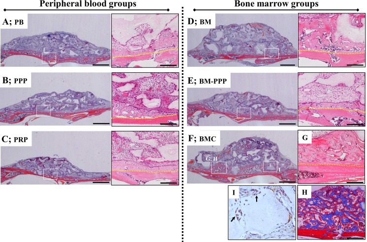
Fig 3. Typical histological appearance in specimens at 2 weeks following transplantation onto the cranium.
Coronal plane sections were stained with hematoxylin and eosin (H&E). For each group, the left panel shows the whole area of specimens (scale bars represent 1 mm), and the right panel shows the magnified image of the white box in the left panel (scale bars represent 200 μm). A small amount of new bone was detected along the host bone in the (A) PB, (B) PPP, (C) PRP, and (E) BM-PPP groups. The (D) BM and (F) BMC groups showed obvious bone formation along the host bone. (G) New bone area surrounded the β-TCP particles and connected with the host bone [box area in (F)]. (H) Masson’s trichrome staining showed the immature (blue) and mature (red) bone in the new bone area [box area in (F)]. (I) Human vimentin immuno-staining showed a few positive cells at the surface of newly formed bone adjacent to the β-TCP particles. The yellow dotted line indicates the boundary between the host bone and the specimen.

