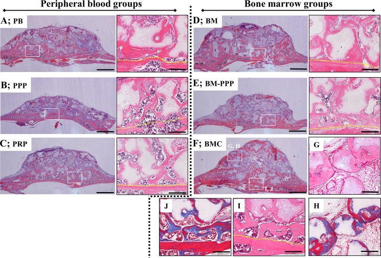
Fig 4. Typical histological appearance in specimens at 4 weeks following transplantation onto the cranium.
Coronal plane sections were stained with hematoxylin and eosin (H&E). For each group, the left panel shows the whole area of specimens (scale bars represent 1 mm), and the right panel shows the magnified image of the white box in the left panel (scale bars represent 200 μm). New bone area was obviously promoted along the host bone and β-TCP particles in the (A) PB, (B) PPP, (C) PRP, (D) BM, (E) BM-PPP, and (F) BMC groups, and replacement bone tissue was clearly observed at the surface of the β-TCP particles in the magnified areas. (G, H) The surface of β-TCP particles was being resorbed and replaced with new bone at the far site from the host bone, and newly formed bone was observed to be more mature (red stained by Masson’s trichrome) [box area in (F)]. (I, J) The newly formed bone was sufficiently integrated with the host bone, and appeared mature (red stained by Masson’s trichrome) [box area in (F)]. The yellow dotted line indicates the boundary between the host bone and the specimen.

