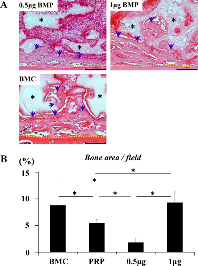
Fig 6. Synergistic effect of BMC and BMP-2 on bone formation.
(A) Bone formation at 2 weeks post-transplantation. Scale bars represent 200 μm. The newly formed bone was integrated with the host bone. rhBMP-2 at 0.5 μg induced a limited amount of new bone formation while 1 μg rhBMP-2 and BMC (BMC represents the BMC group) promoted bone formation to surround β-TCP particles and connect to the host bone. Black asterisk: β-TCP particles, blue arrow: newly formed bone, and yellow dotted line: boundary between the host bone and specimen. (B) Comparison of the area (%) of newly formed bone in each group. PRP represents the PRP group. Values are the means ± standard deviations of five sections from each of the five specimens per group. The asterisk represents statistical significance (*p <0.05) among groups.

