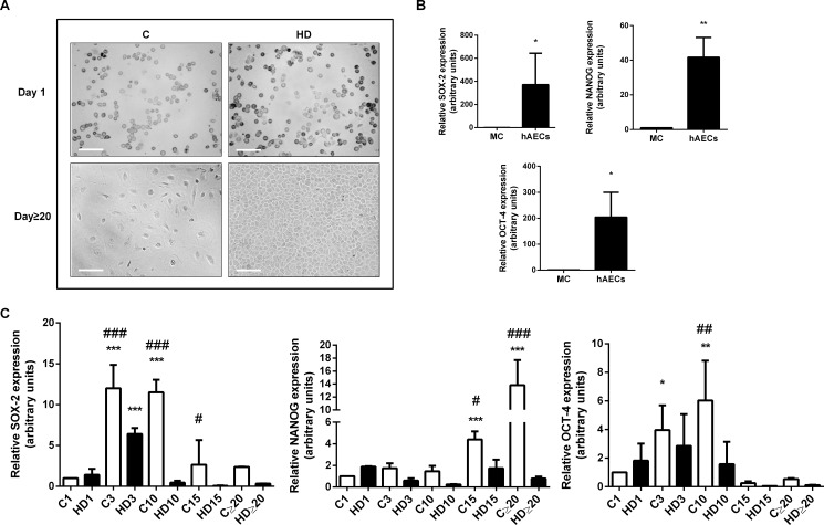
Fig 1. HAECs express pluripotency markers and they diminish during hepatic differentiation.
(A) Amniotic epithelial cells (hAECs) were incubated during 30 days in control medium (C) or treated with hepatic differentiation medium (HD). Representative bright field microscopy images (days 1 and 20) from five independent experiments, at 10X are shown. Scale Bar: 30 μm. (B) Isolated hAECs (1 x 10⁵ cells) were plated in complete IMDM medium supplemented with 10% FBS and incubated during 24 h before RNA extraction. RNA from HepG2 cells (Mature cells = MC) was used as negative control expression. (C) hAECs were incubated with IMDM 10% FBS (C) or with hepatic differentiation medium (HD) for the indicated times (1,3, 10, 15 and more than 20 days) before RNA extraction. In (B) and (C), total RNA was extracted as described in Materials and Methods. SOX-2, OCT-4 and NANOG mRNAs were measured by quantitative real time PCR. CYCLOPHILIN and GAPDH were used as internal standards. Results from a representative experiment are shown and expressed as means ± S.D. for five independent experiments performed in duplicates. *p<0.05, **p<0.01 vs. control day 1; ##p<0.01 vs. respective control.

