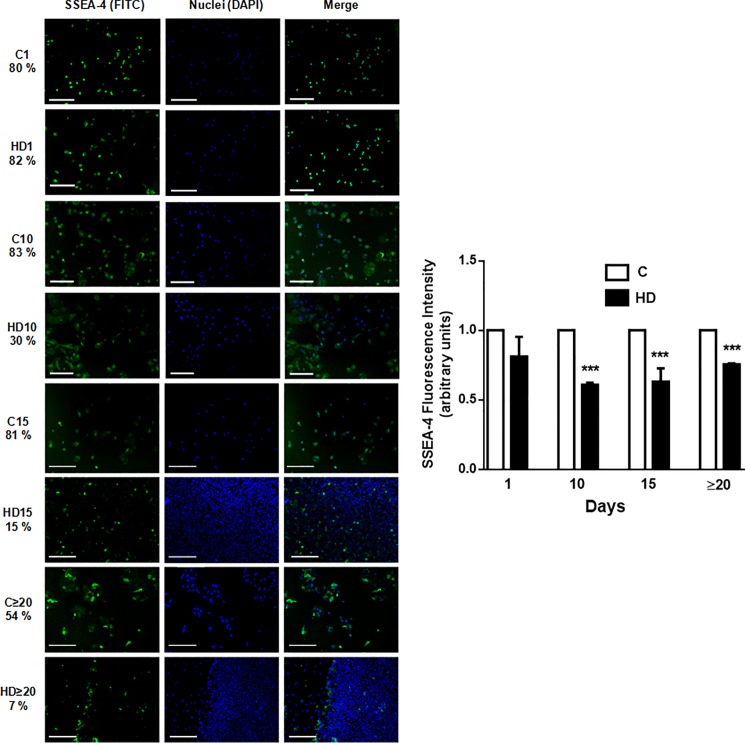
Fig 2. Embryonic antigen SSEA-4 diminishes during HD of hAECs.
Amniotic cells were seeded in 24-wells plate and incubated in IMDM 10% FBS (C) or hepatic differentiation medium (HD), during indicated times. At each time cells were fixed and SSEA-4 expression (green) was detected using FITC conjugated secondary antibody. Representative micrographs from hAECs at different HD times at 10X are shown. The nuclei were stained with DAPI (blue). Percentage of positive cells is shown on the left of images. Graph on the side shows cells fluorescence intensity for SSEA-4. Scale bar: 30 μm. Representative results from three replicates are shown. ***p<0.001 vs. respective control.

