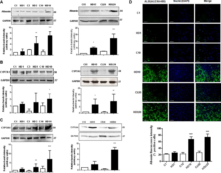
Fig 4. Hepatic proteins expression augments in hAECs with HD treatment.
(A) (B) (C) Amniotic epithelial cells were plated in completed IMDM medium with 10% FBS (C) or hepatic differentiation medium (HD) and incubated during at least 30 days. The medium was changed every three days. At indicated times, cell extracts were prepared and proteins were separated on SDS-PAGE gels. Albumin (A), CYP7A1 (B), and CYP3A4 (C) expression was determined by Western-blot. Molecular weights were estimated using standard protein markers. Loading controls were performed by immunoblotting the same membranes with anti-GAPDH. Bands densitometry is shown in lower panels. Molecular weight (kDa) is indicated at the right of the blot. Representative results from four replicates are shown. (D) Amniotic cells were seeded in 24-wells plate and incubated in IMDM medium supplemented with 10% FBS (C) or hepatic differentiation medium (HD), during indicated times. At each time cells were fixed and albumin expression (green) was detected using Alexa-488 conjugated secondary antibody. Representative micrographs from hAECs at different HD times at 10X are shown. The nuclei were stained with DAPI (blue). Graph below shows positive cells (fluorescence intensity) for albumin. Scale bar: 60 μm. Representative results from three replicates are shown. ***p<0.001 vs. respective control. *p<0.05, **p<0.01, ***p<0.001 vs. control; #p<0.05, ##p<0.01 vs respective control.

