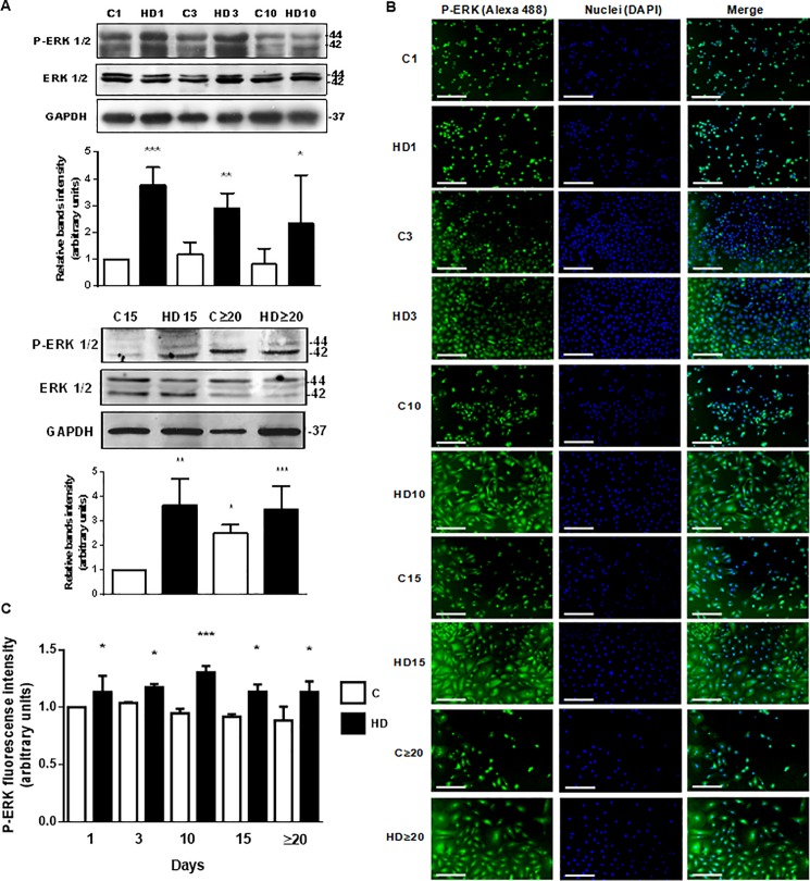
Fig 7. ERK 1/2 phosphorylation is stimulated during hAECs hepatic differentiation.
(A) Amniotic cells were incubated during 1, 3, 10 (top), 15 and up to 30 days (bottom) with IMDM medium 10% FBS (C) or hepatic differentiation medium (HD), as indicated. Extracts from cells were prepared as previously described and loaded in a 12% SDS-PAGE. ERK 1/2 phosphorylation was determined by Western-blot. Loading controls were performed by immunoblotting the same membranes with anti-ERK 1/2 and anti-GAPDH. Bands densitometry is shown in lower panels. Molecular weight (kDa) is indicated at the right of the blot. (B) Amniotic cells were seeded in 24-wells plate and incubated in IMDM medium supplemented with 10% FBS (C) or hepatic differentiation medium (HD), during indicated times. At each time cells were fixed and ERK 1/2 phosphorylation (green) was detected using Alexa-488 conjugated secondary antibody. Representative micrographs at 10X from hAECs at different HD times are shown. The nuclei were stained with DAPI (blue). (C) Graph shows P-ERK fluorescence intensity in hAECs. Scale Bar: 30 μm. Results are expressed as mean ± S.D. for five independent experiments. *p< 0.05, **p< 0.01, ***p< 0.001 vs. control.

