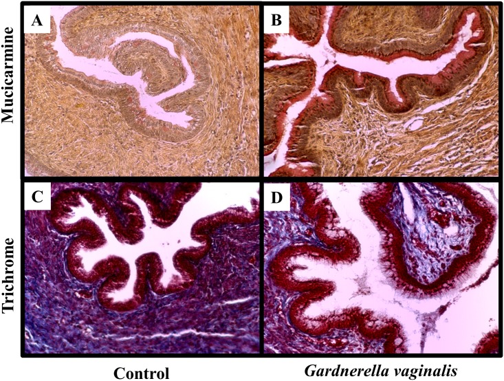
Fig 5. G. vaginalis colonization increased expression of mucin and decreased collagen dispersion within cervical tissues.
Representative cervical sections from Control (A, C) or G. vaginalis (B, D) treated animlas (N = 4 in each group). Cervices were stained with mucicarmine (A, B) for analysis of mucin production while trichrome stain (C, D) shows collagen dispersion. Pictures were taken at a 10X magnification. Trichrome stains collagen blue, muscle fibers red and nuclei black-blue. Mucicarmine stains mucin pink/red, the nuclei blue and any other tissue component yellow.

