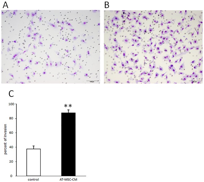
Fig 3. Effects of canine AT-MSC-CM on invasive ability of canine HCC cells.
(A, B) Representative images of Transwell assays observed under a light microscope in the cultures without (A) and (B) with AT-MSC-CM. (C) The percent of invading HCC cells was significantly increased in cells cultured with AT-MSC-CM compared with control cells cultured without AT-MSC-CM. Data are expressed as mean ± SD. **P < 0.01.

