Figure 1. Primary cutaneous B-Cell lymphomas. Dermoscopic features.
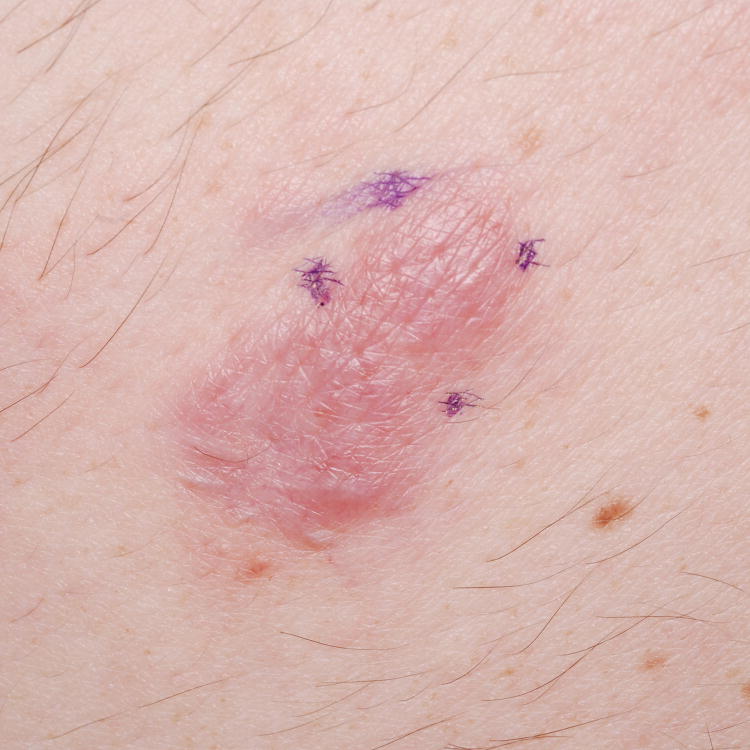
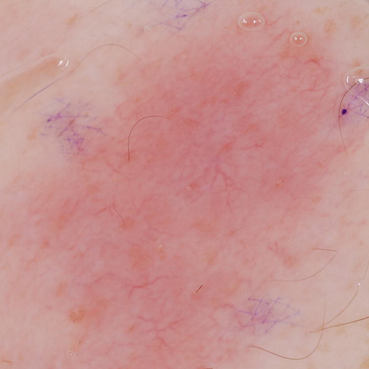
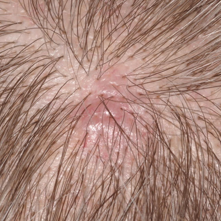
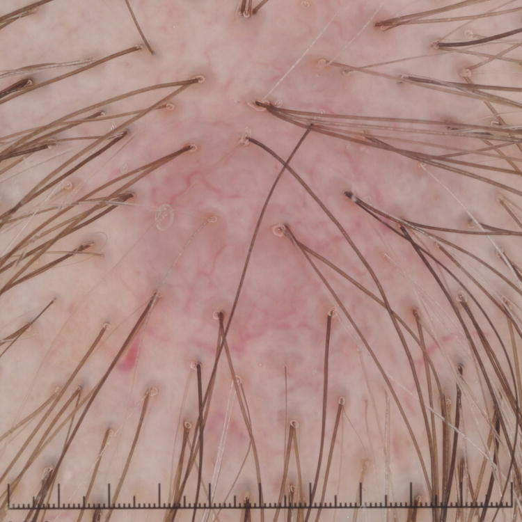
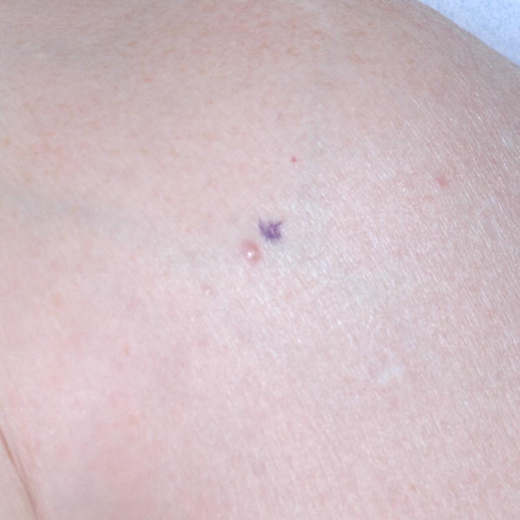
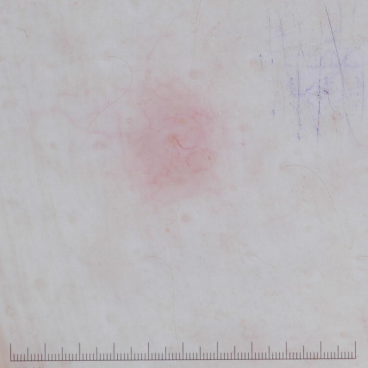
A, Primary cutaneous marginal zone lymphoma presenting as a plaque located on the back. B, Dermoscopic examination reveals salmon-pink-to-orange background with numerous fine, out-of-focus serpentine vessels that are absent from the peri-lesional skin (original magnification, ×10). C, Scalp nodule of primary cutaneous follicle center lymphoma. D, Dermoscopic image showing serpentine vessels within salmon-pink homogeneous area (original magnification, ×10). E, Primary cutaneous follicle center lymphoma presenting as a shiny pink papule on the shoulder. F, Dermoscopic examination reveals few fine, out-of-focus serpentine vessels with salmon-pink to yellowish homogeneous area (original magnification, ×10).
