
Figure 13.
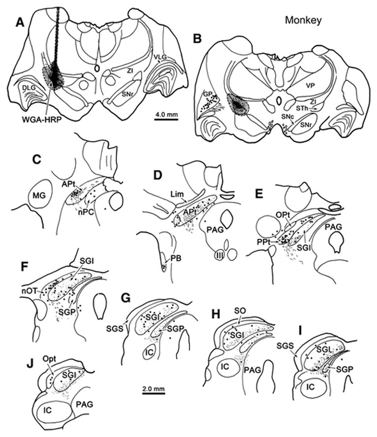
Incertotectal termination in the monkey midbrain tectum. Chartings show the distribution of labeled cells (dots), axons (lines) and terminals (stipple) in the SC (E–J) and pretectum (C–F) following an injection of WGA-HRP into ZI (A–C) in a macaque monkey. Most of the terminal labeling is found in the intermediate gray layer (SGI) and deep gray layer (SGP). Labeled cells were scattered in these layers, as well as in stratum opticum (SO). Labeled terminals and cells were present in the anterior pretectal nucleus (APt)(C&D), posterior pretectal nucleus (PPt)(E), nucleus of the posterior commissure (nPC) and the nucleus of the optic tract (nOT)(F).