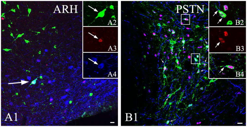
Figure 7.
Connections between the POMC neurons of the arcuate hypothalamic nucleus and the CEA. Confocal microscopic image (A) of triple-immunolabeled section indicates the presence of CTB (green) in a refeeding-activated (c-Fos-positive, red), POMC-producing (blue) perikarion in the arcuate hypothalamic nucleus of brain previously injected with CTB into the CEA. Arrow points to a triple-labeled neuron. The image was made by projecting 7 consecutive 1.8 μm thick confocal optical slices into one plane. To facilitate identification of triple-labeled cells, CTB, c-Fos and POMC-labeling of the same cells are shown in separate images in the insets. Note that the vast majority of the c-Fos-containing POMC neurons do not contain CTB-immunoreactivity.
Confocal image of a triple-immunolabeled section (B) shows POMC-IR (blue) boutons on refeeding-activated neurons (c-Fos-IR, red) in the parasubthalamic nucleus (PSTN) that projects to the CEA (CTB-IR, green). Arrowheads point to POMC-IR varicosities on the surface of double-labeled, c-Fos-and POMC-IR neurons. The cells in squares are shown in higher magnification in the insets. B3 shows the c-Fos-IR nucleus of the neuron illustrated in B2. The POMC-IR varicosities contacting the perikaryon and the dendrite of this double-labeled neuron are pointed by arrows. B3 also illustrates a refeeding-activated and CEA-projecting neuron contacted by a POMC-IR varicosity (arrow). Note that primary antibodies against c-Fos and POMC were produced in rabbit, thus immunolabeling was performed sequentially, which prevented cross-reaction in the cytoplasm, but not in the cell nuclei. Scale bars: 20μm.

