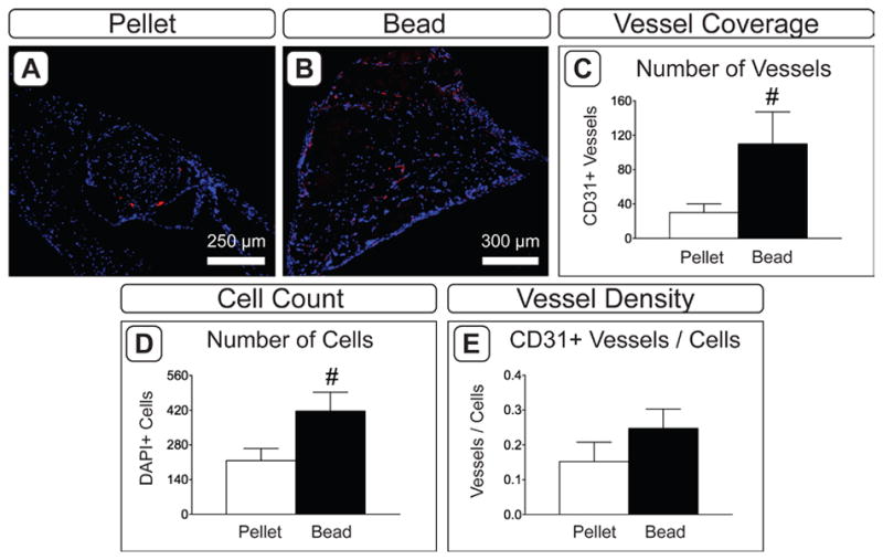
Figure 5.

Vascular invasion in 8 week explants. A,B) CD31 immuofluorescence in pellet (A) and bead (B) constructs. Red indicates positive staining; blue indicates cell nuclei (DAPI). C–F) Assessment of vascularization in explanted tissues. Blood vessel coverage was assessed by quantifying the number of CD31+ vessels (C) and the number of cell nuclei (D). A representative value for blood vessel density was calculated by normalizing to the number of vessels to the number of DAPI-positive cells (E). (n = 6; #: p < 0.065).
