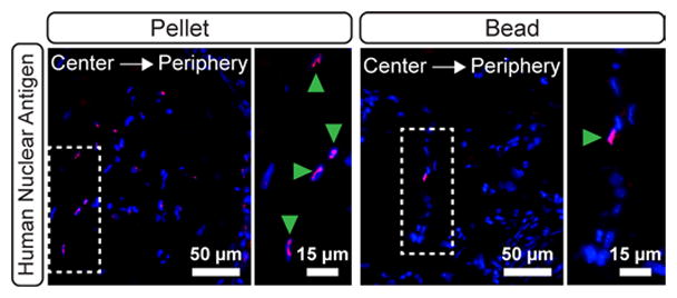
Figure 6.

Contribution of hASCs in 8 week explanted pellets (A) and beads (B). Magenta staining indicates positive staining for human nuclear antigen on hASCs. Blue indicates all cell nuclei (DAPI). The white dashed boxes indicate the locations of magnified areas in the right-hand panels. Green arrowheads denote human cells in the magnified regions.
