Fig. 2.
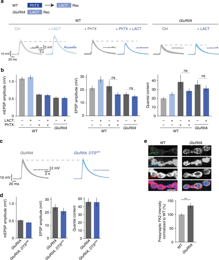
Proteasome perturbation does not potentiate release after acute induction or sustained expression of PHP. a Scheme for treatment of NMJs and representative EPSP and mEPSP traces. Wild-type NMJs were incubated with saline or 20 µM PhTX for 10 min, followed by a wash and treatment with saline or 100 µM lactacystin for 30 min at 0.3 mM [Ca2+]e. GluRIIA-mutant synapses were treated with saline or 100 µM lactacystin for 30 min at 0.3 mM [Ca2+]e. b Quantification of EPSP amplitude, mEPSP amplitude and quantal content for larvae treated as described in (a). Lactacystin application to larvae treated with PhTX or to GluRIIA-mutant larvae has no effect on EPSP amplitude or quantal content. Mean ± s.e.m.; n ≥ 10 cells; ns: not significant; Student’s t-test. c Representative EPSP and mEPSP traces of GluRIIA-mutant synapses and GluRIIA,DTSpre double-mutant synapses. d Quantification of mEPSP amplitude, EPSP amplitude and quantal content of the genotypes described in (c). Presynaptic proteasome perturbation does not increase EPSP amplitude or quantal content in GluRIIA-mutant synapses. Mean ± s.e.m.; n ≥ 7 cells. e) Representative NMJs stained for mono- and polyubiquitinated proteins (aFK2), neuronal membrane (aHRP, horseradish peroxidase), and postsynaptic reticulum (aDLG, discs large) of wild-type and GluRIIA-mutant larvae (scale bar, 5 µm). Quantification of FK2-fluorescence intensity in the HRP mask (presynapse) suggests that GluRIIA-mutant NMJs have higher levels of mono- and polyubiquitinated proteins compared to wild-type NMJs. Mean ± s.e.m.; n = 24 NMJs; **p < 0.001; Student’s t-test

