Figure 4.
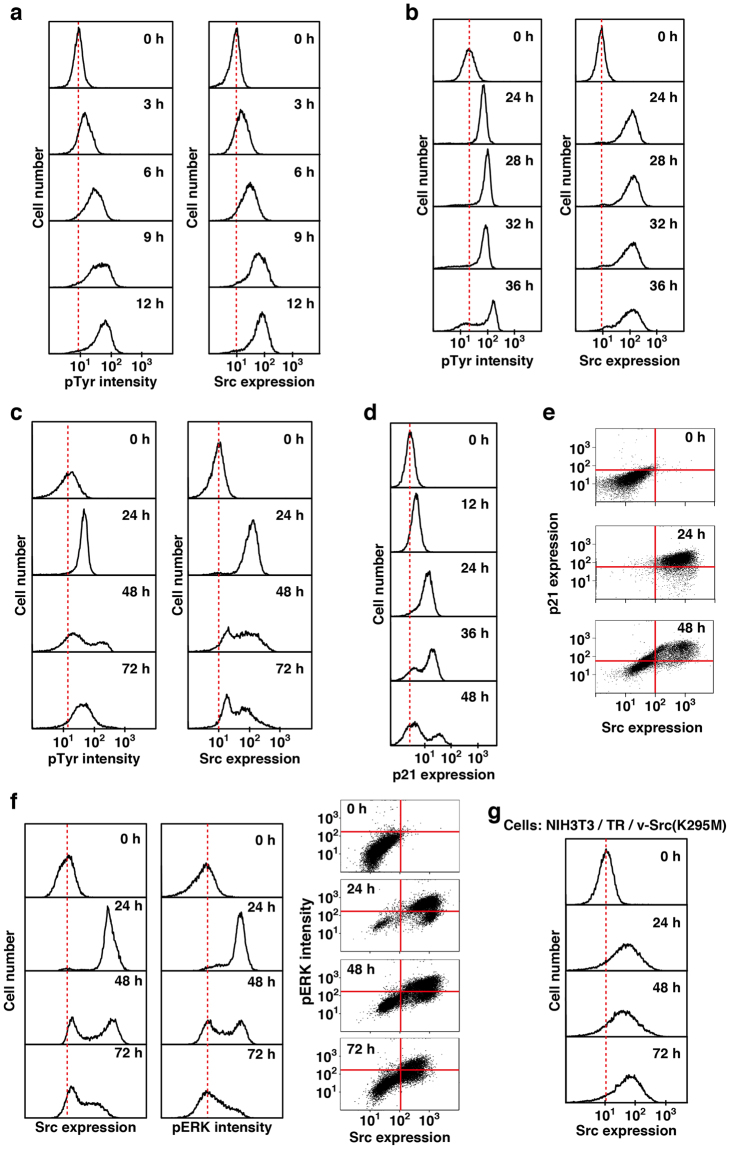
Suppression of v-Src expression in NIH3T3 cells. (a–f) NIH3T3/TR/v-Src-wt cells were treated with 1 µg/ml Dox for the indicated times. (a–c) Cells were fixed and doubly stained with anti-Src (#327) and anti-pTyr antibodies for flow cytometry. (d) Cells were fixed and stained with anti-p21 antibody for flow cytometry. (e) Cells were fixed and doubly stained with anti-Src (#327) and anti-p21 antibodies for flow cytometry. Two-dimensional histograms (Src vs p21) are presented. (f) Cells were fixed and doubly stained with anti-Src (#327) and anti-pERK1/2 antibodies for flow cytometry. Src and pERK histograms (left) and two-dimensional histograms (Src vs pERK) (right) are presented. (g) NIH3T3/TR/v-Src(K295M) cells were treated with 1 µg/ml Dox for the indicated times. Cells were fixed and stained with anti-Src (#327) antibody for flow cytometry.

