Figure 6.
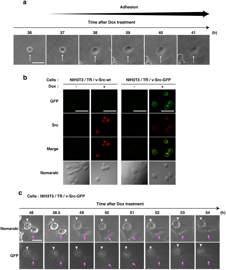
Suppression of v-Src expression in re-adherent cells. (a) Living NIH3T3/TR/v-Src-wt cells were monitored by time-lapse phase-contrast microscopy from 36 h after Dox addition. Arrows indicate a cell that had been detached by v-Src expression and re-adhered to a culture dish. Scale bar, 50 µm. (b) NIH3T3/TR/v-Src-wt cells and NIH3T3/TR/v-Src-GFP cells were cultured for 24 h with or without 1 µg/ml Dox. Expressed proteins were visualized with anti-Src (#327) antibody (red) and GFP fluorescence (green). Scale bars, 50 µm. (c) Living NIH3T3/TR/v-Src-GFP cells were monitored by time-lapse phase-contrast microscopy from 48 h after Dox addition. White arrowheads indicate a cell that had been detached and continuously expressed v-Src-GFP. Red arrows indicate a cell that had been detached and re-adhered to a culture dish and subsequently exhibited cell spreading while expression of v-Src-GFP was suppressed. Scale bar, 25 µm.

