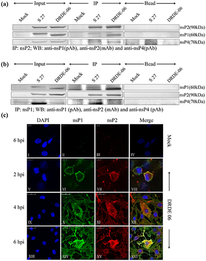
Figure 1.
CHIKV nsP2 interacts with nsP1 during virus infection in Vero cells through RC. (a) Vero cells grown in 100 mm dish, were either infected with S 27 or DRDE-06 virus at an MOI of 2. Mock infected cells were considered as negative control. The cells were harvested at 6 hpi and 10 hpi for DRDE-06 and S 27 respectively. Immunoprecipitation was performed with the lysates using CHIKV anti-nsP2 mAb and Western blot was carried out using anti-nsP2 mAb, anti-nsP1 and anti-nsP4 pAbs. Beads with normal IgG serve as negative control. (b) Same Vero cell lysates were immunoprecipitated with anti-nsP1 pAb and elutes were separated in 10% SDS-PAGE. Western blot was performed using anti-nsP1 pAb, -nsP2 mAb and -nsP4 pAb. (c) Vero cells were plated onto cover-slips and infected either without virus (mock) or with DRDE-06 at a MOI of 2. The cells were fixed after 2, 4 and 6 hpi, probed together with anti-nsP1 pAb (II, VI, X, XIV) and anti-nsP2 mAb (III, VII, XI, XV) followed by staining with secondary antibodies, anti-rabbit Alexa Fluor 488 (green) or anti-mouse Alexa Fluor 594 (red) respectively. Nuclei were counterstained with DAPI (blue). Fluorescent images were acquired using the Leica TCS SP5 confocal microscope.

