Fig. 5.
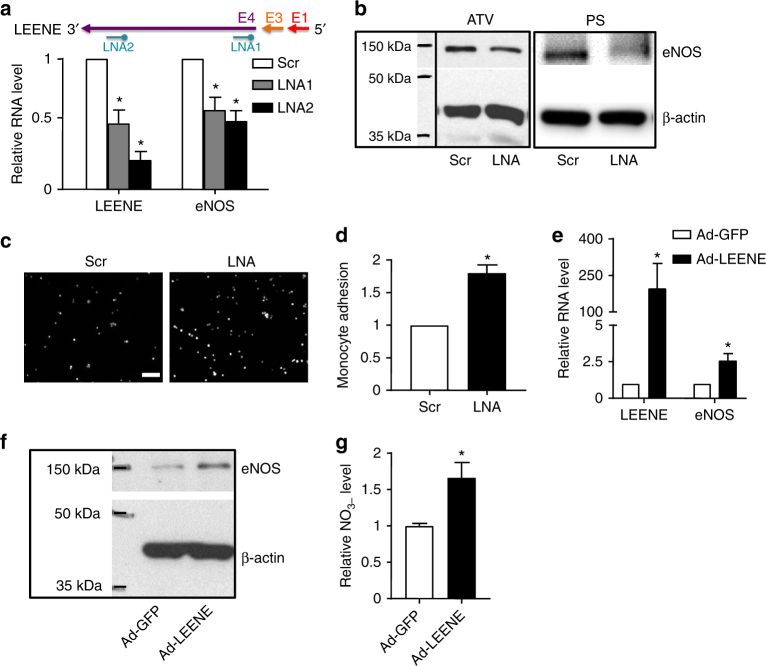
LEENE RNA regulates eNOS expression and EC function. a–e HUVECs were transfected with LNA (50 nM) targeting Exon 4 of LEENE. Basal RNA levels of LEENE and eNOS were detected by qPCR in a. Protein levels of eNOS in HUVECs treated with ATV or PS were revealed by immunoblotting. c, d ECs were transfected with scramble or LEENE LNA before subjected to PS for 12 h. Fluorescence-labeled THP-1 cells were added to the EC monolayer, and the monocytes adhering to ECs were visualized by fluorescence microscopy (scale bar = 100 μm). The representative images are shown in c and the quantification based on five randomly selected fields per group per experiment are shown in d. e–g HUVECs were infected with Ad-GFP or Ad-LEENE for 48 h. RNA levels of LEENE and eNOS were detected by qPCR (e), protein level of eNOS in HUVECs was revealed by immunoblotting (f), and NO production was measured by a fluorometric assay (g). Densitometry analysis of immunoblotting shown in b and f was performed (Supplementary Fig. 9). Data are presented as mean ± SEM. n = 3–5 in each group. Student’s t test was used. *p < 0.05 compared to scrambled control or Ad-GFP in respective experiments

