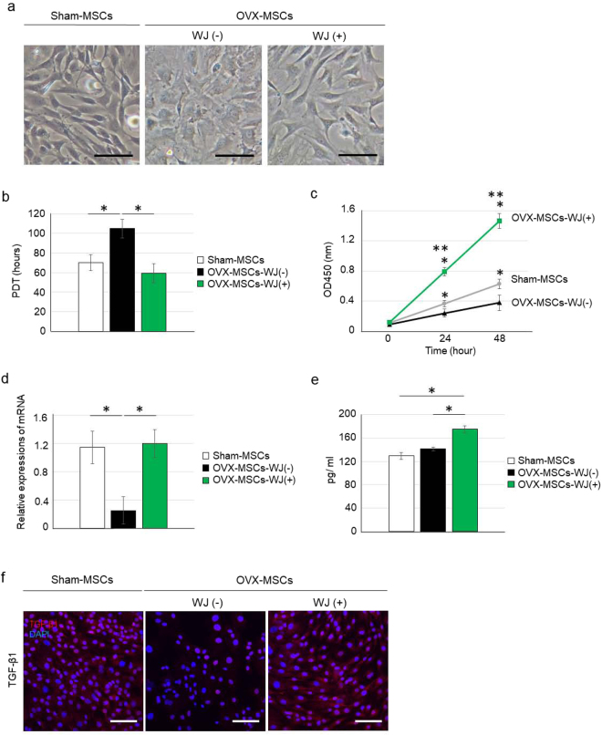
Figure 2.
Abnormalities and activating effects of WJS for cell morphology, proliferation, secretion, and protein expression of OVX-MSCs. (a) Phase contrast observations of Sham-MSCs (left panel), OVX-MSCs-WJ(-) (middle panel) and OVX-MSCs-WJ(+) (right panel). Images were obtained at 48 h after activation with WJS. Bar: 100 μm. (b) Population doubling time of Sham-MSCs, OVX-MSCs-WJ(-), and OVX-MSCs-WJ(+) at passage 2. *P < 0.05. Data are expressed as mean ± SE of 5–6 BM-MSC cultures. (c) MTT proliferation assay of Sham-MSCs, OVX-MSCs-WJ(-), and OVX-MSCs-WJ(+). *P < 0.05. Data are expressed as mean ± SE of 5–6 BM-MSC cultures. (d) Relative mRNA expression of Sham-MSCs, OVX-MSCs-WJ(-), and OVX-MSCs-WJ(+). *P < 0.05. Data are expressed as mean ± SE of 4 BM-MSCs. Opg, osteoprotegerin. (e) OPG levels in the culture supernatant of Sham-MSCs, OVX-MSCs-WJ(-), and OVX-MSCs-WJ(+). *P < 0.05. Data are expressed as mean ± SE of 4 BM-MSC cultures. (f) Immunofluorescence staining of Sham-MSCs (left panel), OVX-MSCs-WJ(-) (middle panel), and OVX-MSCs-WJ(+) (right panel) with an anti-TFG-β antibody (red). DAPI was used for counterstaining nuclei (blue). Bar: 100 μm.

