Figure 5.
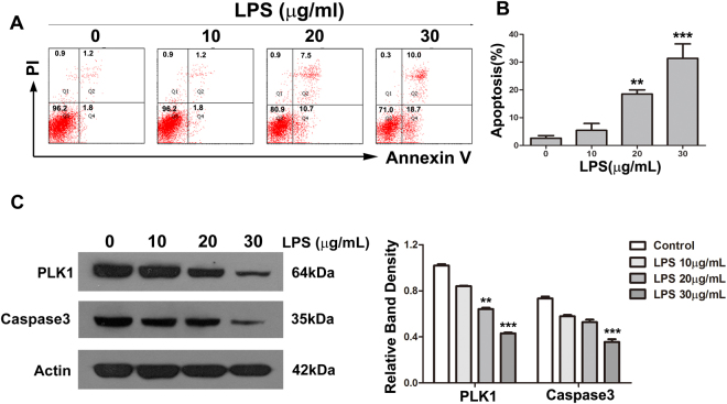
LPS induces apoptosis in HT29 cells. (A) HT29 cells were exposed to various concentrations of LPS for 24 h. Apoptosis was analysed by Annexin V-FITC/PI double-labelling assay. (B) The degree of apoptotic cell death was quantified. Data represent the mean ± SD (**P < 0.01, ***P < 0.001 compared with control group.). (C) The levels of PLK1 and caspase-3 in HT29 cells after treatment with LPS at various concentrations for 24 h. The graph represents the relative band densities. Values are mean ± SEM (n = 3). **P < 0.01, ***P < 0.001 versus control group.

