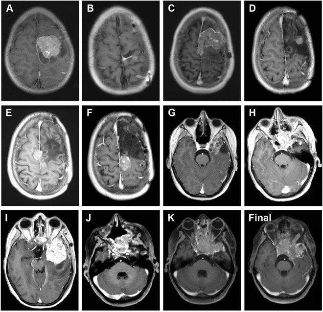
Figure 3.
Post-contrast, T1 weighted axial MRI images of all recurrences. (A–K) representative images prior to 1st to 11th surgical resections respectively. (L), representative image of final tumour progression. (A) age 6.79, underwent surgical resection of left frontal tumour. (B) age 7.33, left frontal craniotomy, partial resection, treated with radiotherapy and temozolomide. (C) age 9.11, left frontal subtotal resection, treated with CCNU and temozolomide. (D) age 11.26, biopsy. (E) age 13.10, resection. (F) age 14.04, resection. (G) age 14.29, right temporal craniotomy, gross total resection. (H) age 14.67, sphenoid tumour, endoscopic endonasal sphenoidectomy. (I) age 14.96, left temporal resection. (J) age 16.05, nasal biopsy. (K) age 16.17, nasal cavity resection. The patient died aged 16.42 years (Final).

