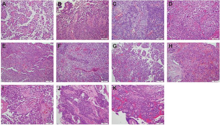
Figure 4.
Histopathological features of all recurrences. (A) Initial presentation – age 6.79 years (as in Fig. 1). (B) Similar morphology with high focal mitotic activity, fewer perivascular pseudorosettes and sharply demarcated borders. Evidence of calcification. (C) More cellular tumour with only single ribbon-like structures and almost no pseudorosettes seen. (D) Similar morphology to C, with hyalinised vessels. (E) Fewer mitotic figures present, and cells seen with conspicuous nucleoli. (F) Similar to (B), but with no calcification. Increased perivascular ribbons and necrotic foci. (G) Distinct lesion with prominent cellular pleomorphism and large hyperchromatic nuclei. Many cells have an epithelioid appearance. There are papillary structures and numerous pseudorosettes. (H) Tumour is infiltrating the sphenoid bone and sinus and has numerous invasive, pleomorphic cells with hyperchromatic and large nuclei. (I) Well vascularised pleomorphic tumour with several pseudorosettes. (J) Tumour was mostly necrotic tumour but viable tissue. showed pseudorosettes around hyalinised vessels. (K) Tumour was mostly necrotic. but preserved tumour shows numerous pseudorosettes, mostly around hyalinised vessels with thick walls. Original magnification x200.

