Figure 2.
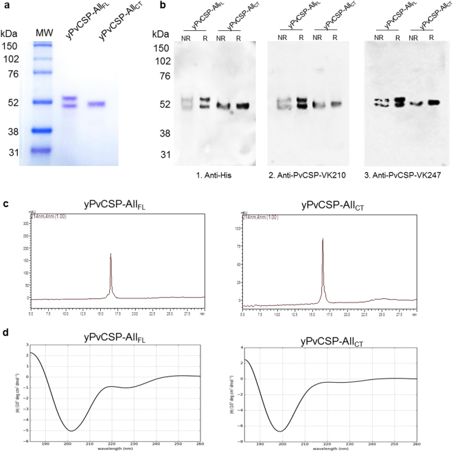
Expression, purification, and biophysical analyses of recombinant proteins. (a) SDS-PAGE (12%) of yPvCSP-AllFL and yPvCSP-AllCT proteins, stained with Coomassie blue under reducing conditions. (b) Immunoblotting analyses of recombinant yPvCSP-AllFL and yPvCSP-AllCT. (1). Anti-histidine tag mAb (diluted 1:1,000, GE Healthcare); (2). Anti-PvCSP-VK210 mAb (diluted 1:1,000) 3. Anti-PvCSP-VK247 mAb (diluted 1:11,000). NR and R: proteins under non-reducing and reducing conditions (with 2-mercaptoethanol), respectively; (c) The purity of proteins yPvCSP-AllFL and yPvCSP-AllCT, after a combination of chromatographic separations, was analysed by RP-HPLC. The gradient elution was developed with 0.1% TFA in water and 0.1% TFA in 90% acetonitrile at 22 °C, and a rate of 1 mL/min for 30 minutes on a C18 column (Phenomenex). MW: molecular weight. (d) The secondary structure of yPvCSP-AllFL and yPvCSP-AllCT was analysed by a circular dichroism (CD) spectrum. The CD spectrum of the recombinant proteins was recorded from 190 to 260 nm using a JASCO-J815 spectropolarimeter.

