Fig. 1.
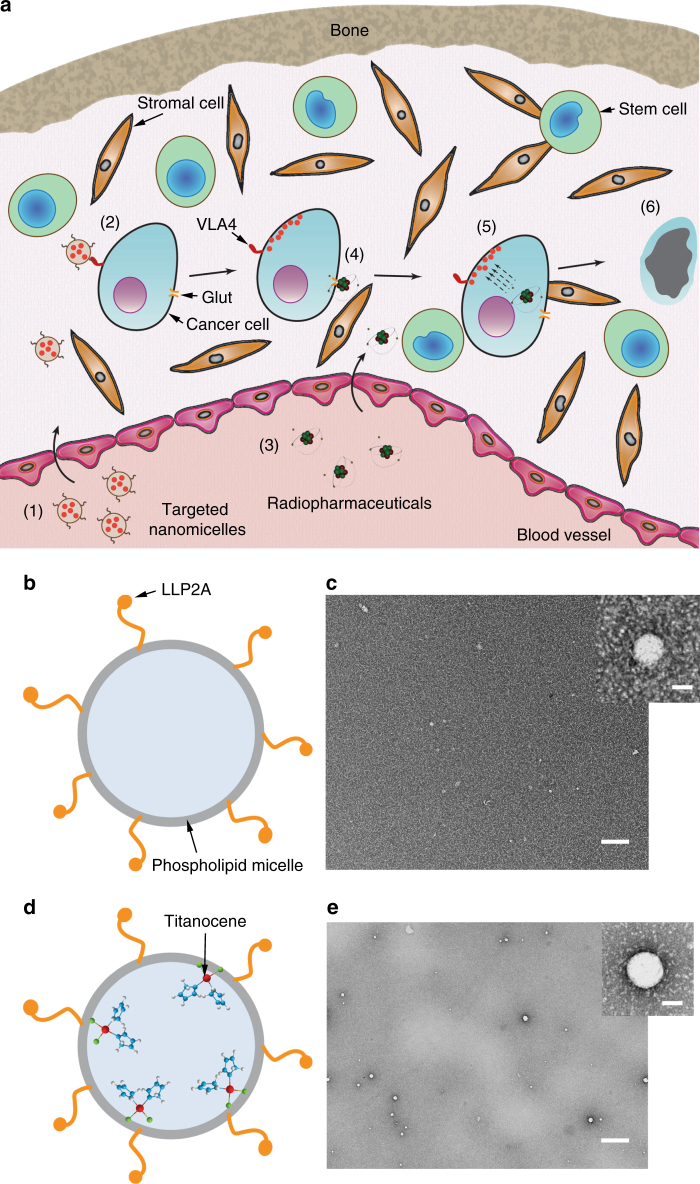
Orthogonal cancer targeting strategy using nanomicelles. a Schematic of the process of photoactivation of Titanocene in disseminated cancer cells in the bone marrow microenvironment. The various phases are numbered: 1. Administration of targeted NM-TC; 2. The targeted NM enter the bone marrow from the vasculature and bind to α4β1 receptor on the cancer cells and subsequently deliver the drug to the cell; 3. Administration of radiopharmaceuticals (18FDG), which is typically 1.5–2 h after phase (1); 4. 18FDG enters the cancer cells through the overexpressed Glut transporters on cancer cells; 5. Once the drug and radiopharmaceutical are co-localized in the cancer cells, the former is photoactivated by the latter through CR leading to cell death (6). Notice that since the other vital cells in the bone marrow, such as stem cells and stromal cells, do not express the combination of α4β1 and glut receptors essential for the treatment to work, they would largely remain unaffected causing minimal off-target toxicity. b Schematic of phospholipid NM with VLA-4 homing ligands. c TEM image of micelles alone. Scale bar, 100 nm. Inset: single micelle. Scale bar, 10 nm. d Schematic of phospholipid NM encapsulating TC with VLA-4 homing ligands. e TEM image of micelle incorporated with TC in the membrane. Scale bar, 100 nm. Inset: single NM-TC. Scale bar, 10 nm

