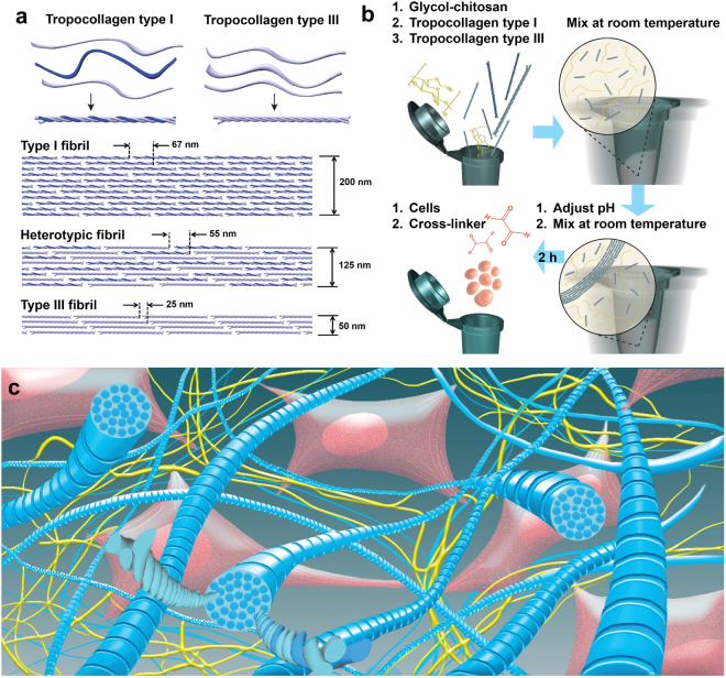
Figure 1.
(a) Schematic of tropocollagen types I and III followed by their arrangements to form type I fibrils, heterotypic fibrils of types I and III (I&III), and type III fibrils. These illustrations are further supported by data reported in a recent study, in which average (fibril diameter, periodicity) of (200,67), (125,55) and (50,25) were obtained for types I, I&III with a mixing ratio of 1:1, and III fibrils, respectively23; (b) Schematic of the step-by-step fabrication procedure. Tropocollagen types I and III molecules were added to glycol-chitosan (GCS) solution, and the mixture was vortexed at room temperature. After adjusting pH to the physiological pH level, the mixture was vortexed again. At this stage, the mixture includes both tropocollagen molecules and newly-formed collagen fibrils. After 2 hours, cells were added and properly mixed. Finally, the cross-linker (glyoxal) was added, and the mixture was mixed to ensure a homogenous cell distribution; (c) Schematic of the three-dimensional structure of the nano-fibrillar hybrid hydrogel (Col-I&III/GCS). Heterotypic collagen fibrils (shown in blue) were randomly distributed in GCS matrix (shown in yellow). Heads of the tropocollagen molecules are shown on the cross-sections of the representative fibrils. Glyoxal was used to form covalent cross-linking between GCS molecules as well as between collagen fibrils and GCS matrix. The proposed hydrogel supports cell adhesion because of cell attachments to collagen fibrils, as illustrated (Col-I&III: the simultaneous presence of Col-I and Col-III).

