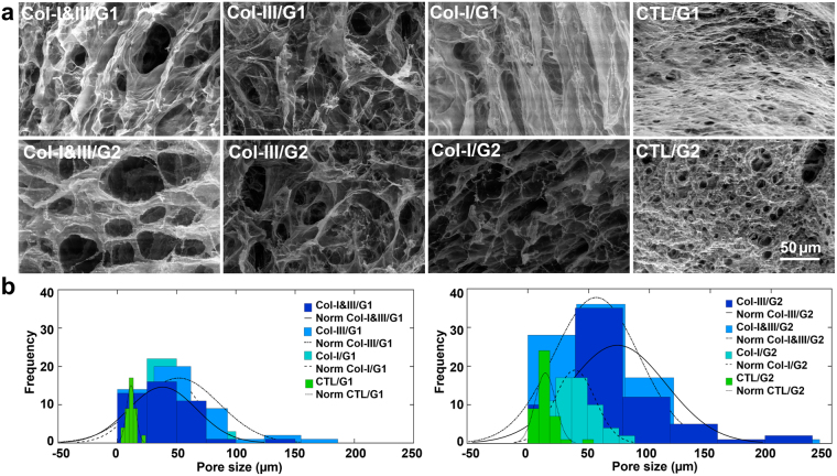
Figure 3.
Environmental scanning electron microscopy (ESEM) results. (a) Representative ESEM images of Col-I&III/glycol-chitosan (GCS), Col-III/GCS, positive control (Col-I/GCS) and negative control (CTL/GCS) hydrogels. Porous structures with interconnected pores of irregular shapes were found for the Col/GCS hydrogels. Rounded closed pores with smaller dimensions were found for the negative controls. The pore size increased with the incorporation of collagen in GCS hydrogels; (b) Histograms of the measured pore sizes out of three replicates for each study group. The mode was slightly larger for Col-III/GCS hydrogels, compared with those of associated Col-I&III/GCS hydrogels and positive controls (Col-I/GCS), which were all greater than those of the associated negative controls (Col-III: collagen type III, Col-I: collagen type I, Col-I&III: the simultaneous presence of Col-I and Col-III, and CTL: negative controls. G1 and G2 represent the final GCS concentration of 2% and 1%, respectively).

