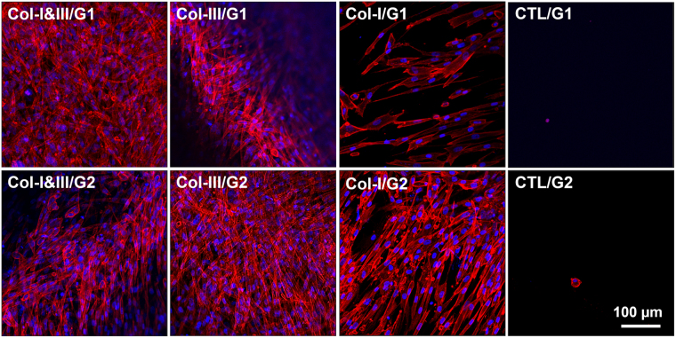
Figure 5.
Representative fluorescent images of adherent cells cultured on the surface of the collagen (Col)/glycol-chitosan (GCS) hydrogels and negative controls. Actin cytoskeleton and nuclei are shown in red and blue, respectively. Fibroblasts were attached strongly to the surface of the proposed Col/GCS hydrogels. The cells were spread out extensively, and the formation of actin stress fibers were observed. On the contrary, only a few rounded cells were found attached on the surface of the negative controls after 3 days. The images were captured with a 20x objective, and the scale bar shows 100 μm (Col-III: collagen type III, Col-I: collagen type I, Col-I&III: both Col-I and Col-III, and CTL: negative controls. G1 and G2 represent the final GCS concentration of 2% and 1%, respectively).

