Fig. 1.
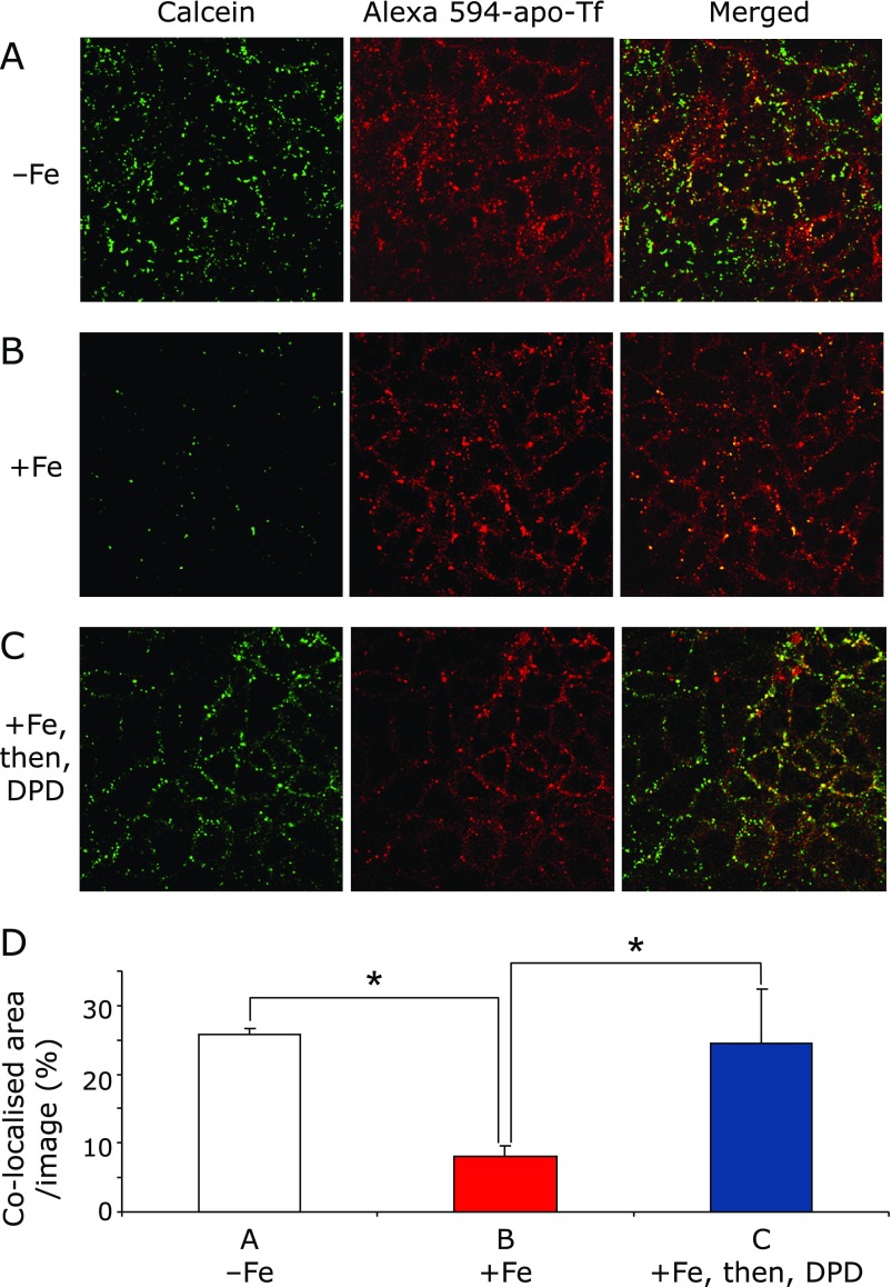
Co-localization of the metallosensor calcein and apo-transferrin (Tf) were examined in different iron conditions by confocal microscopy. (A) Observation without iron feeding. After calcein and apo-Tf were added into the basal chamber and incubated for 20 min, the Caco-2 monolayer was extensively washed and fixed. (B) Observation with iron feeding. After extensive washing, iron-ascorbate complex (5 µM Fe, 500 µM ascorbate) was added into the apical chamber and incubated for 20 min with the Caco-2 monolayer. (C) Observation of 2,2'-dipyridyl (DPD) addition after iron feeding. After iron feeding, DPD (200 µM) was added into the apical chamber and incubated for 5 min. Shown images are representative of the fluorescent probes. (D) Digital analysis of fluorescent signals. Images, which were collected in (A), (B) and (C) experimental conditions, were analyzed by counting the signal intensity and area in digital pixels by MetaMorph. These analyses are expressed in co-localized area/image (%) mean ± SD for at least three independent experiments. * indicates a significant difference (p<0.05).

