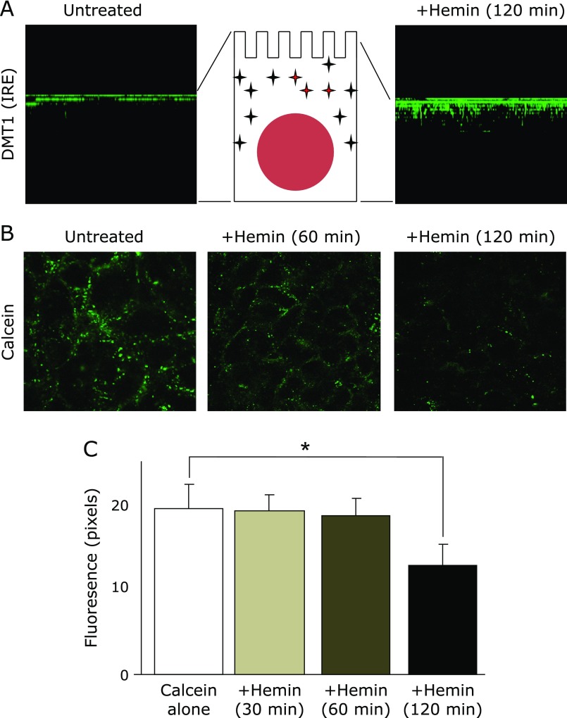
Fig. 3.
Impact of hemin feeding on DMT1 (IRE) internalization and labile iron pools. (A) Immunofluorescent staining of DMT1 (IRE) in the Caco-2 monolayer. (Left) Before feeding, DMT1 (IRE) was localized in the BBM. (Right) After feeding with hemin (50 µM) for 2 h, DMT1 (IRE) migrated into the subapical compartment. These images were reconstructed to be viewed from a lateral perspective by taking sections along the z-axis with a step size of 2 µm to give ~12 sections per imaged field. (B) Determination of the labile iron pools by calcein after hemin feeding. After feeding with hemin (50 µM), Caco-2 monolayers were stained with calcein (200 µM). Quenching of these fluorescences were observed 120 min later but were not significant 30 and 60 min later. (C) Digital analysis of fluorescent signals. Images, which were collected after hemin feeding, were analyzed for signal intensity and area in digital pixels by MetaMorph. These analyses were expressed in fluorescence mean ± SD for at least three independent experiments. * indicates a significant difference (p<0.05).

