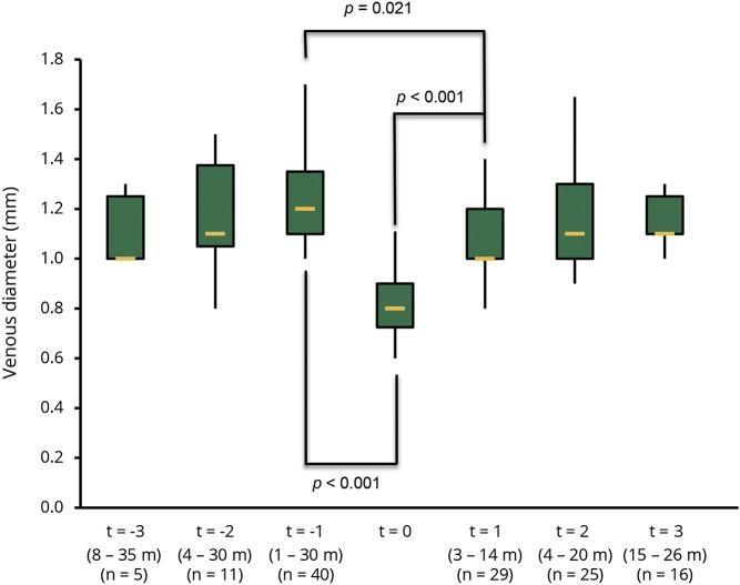
Figure 1. Box plot of the venous diameter in lesions.
Box plot of the venous diameter of 7 MRI examinations in chronological order. X-axis: relative time points for comparison with t = 0, range of intervals as given below in months (m). The number of analyzed veins (n) is given below. Y-axis: venous diameter (mm).

